Technology
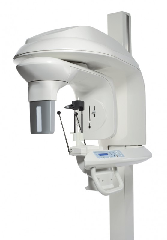
Root Canal Experts is equipped with Cone Beam Computed Tomography (CBCT); this device provides a detailed three-dimensional scan of the tooth being treated. The level of detail provided by this technology ensures a much higher success rate of treatment and eliminates most of the current diagnostic limitations.
Our practice is the first in the area to add a 3D extraoral imaging system to its office, revolutionizing patient treatment and perfecting the way the practice treats oral infections. The KODAK 9000 3D Extraoral Imaging System enables our doctors to obtain low radiation dose, high-resolution, three-dimensional (3D) images, as well as panoramic images.
With the addition of this state-of-the-art 3D unit, Root Canal Experts will greatly improve its level of patient care. Three-dimensional technology allows doctors to better visualize their patients’ dentition, without having to send patients for radiology scans. Viewing an unprecedented level of anatomical detail helps practitioners diagnose more accurately and treat with confidence. The KODAK 9000 3D System will transform dental imaging in the same way that CT scans have changed the medical field, in terms of care through better visualization.
This unique “two-in-one” system (3D and panoramic) is well suited for dental professionals who regularly perform complex diagnostic, restorative, surgical, and endodontic procedures. The highest resolution imaging capabilities provided by this unit will enable our practice to detect lesions with more accuracy. This breakthrough technology provides unprecedented x-ray views of the oral cavity.
Periapical and panoramic radiography have been augmented by the recent introduction of high-resolution cone beam computed tomography (CBCT), allowing 3D assessment of oral lesions, canal morphology, retreatment cases, root fractures, implants, and so forth. The KODAK 9000 3D System uses less radiation than other systems, radiating only one small area of view at a time. Comfortable patient positioning and wheelchair accessibility make this unit patient-friendly. The system enables our practice to perform a wider range of diagnoses and treatments in the office, helping reduce multiple visits, saving patients time and making the treatment more affordable.

Cone Beam Computed Tomography
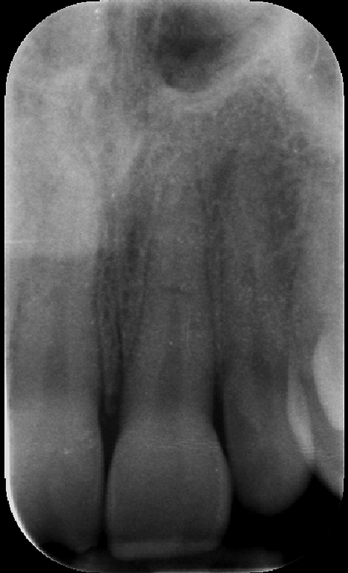
The limitations of radiographs have been well documented, in this case a young patient suffered a traumatic injury and there appears to be a fracture on the radiograph but it’s extent is unclear.

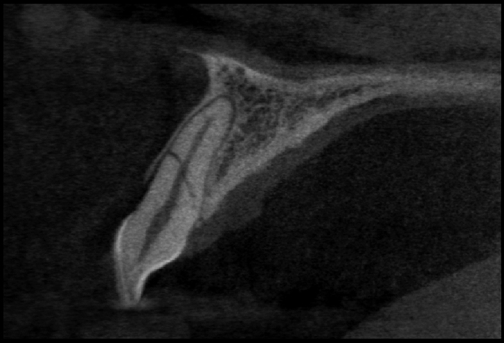
The patient was then taken to have a CBCT scan done at our facility and the scan revealed 2 clear oblique fractures, one at the mid root level and another one at the level of the apical third.
